Voltage Sensitive Dye Data
High Speed Voltage Sensitive Dye Imaging of Spontaneous Activities in Cultured Neuronal Network
- Sample
- Cultured Neurons
- Fluorescence Dye
- FluoVolt
- Imaging System
- MiCAM05-C35IR
- Pixels
- 636x360
- Frame Rate
- 277fps (3.6msec/frame)
Voltage Sensitive Dye Imaging of Brain Slice (MiCAM05-N256)
- Sample
- Brain Slice
- Method
- Electrical stimulation
- Fluorescence Dye
- Voltage sensitive dye (Di-4-Anepps)
- Imaging System
- MiCAM05-N256
- Pixels
- 256x256
- Frame Rate
- 1,000fps (1.0msec/frame)
- Provided by
- Dr. Takashi Tominaga, Institute of Neuroscience, Tokushima Bunri University
Voltage-Sensitive Dye Imaging of Mouse Brain Slice (MiCAM ULTIMA)
- Sample
- Mouse Brain Slice including Cortex and Hippocampus
- Method
- Electrical stimulation
- Fluorescence Dye
- Voltage sensitive dye (Di-4-Anepps)
- Imaging System
- MiCAM05-D225
- Pixels
- 256x240 pixels
- Frame Rate
- 1,000fps (1.0msec/frame)
- Provided by
- Dr. Takashi Tominaga, Institute of Neuroscience, Tokushima Bunri University
Mouse In Vivo Voltage-Sensitive Dye Imaging (MiCAM ULTIMA-H)
This data was obtained from a 2 month-old mouse that was under urethane anaesthesia (1.75 g/kg). A heating pad was used to maintain the measured body temperature of the mouse at 37°C. Two electrodes were inserted under the skin of the forearms to receive the electrocardiogram signal which was used to trigger the MiCAM ULTIMA acquisition.
- Sample
- Mouse In Vivo Bain (Somatosensory Barrel Cortex)
- Method
- Piezo Whisker Flick Stimulation
- Fluorescence Dye
- Voltage sensitive dye (RH-1691)
- Imaging System
- MiCAM ULTIMA
- Pixels
- 100x100 pixels
- Frame Rate
- 500fps (2.0msec/frame)
- Provided by
- Dr.Isabelle Ferezou and Dr.Carl C.H. Petersen, Brain Mind Institute, EPFL, Switzerland
- Reference
- Visualizing the Cortical Representation of Whisker Touch: Voltage-Sensitive Dye Imaging
in Freely Moving Mice.
Neuron. 2006 May 18;50(4):617-29.
Rat In Vivo Optical Mapping using Voltage-Sensitive Dye (MiCAM02-HR)
- Sample
- Rat In Vivo Bain (Insular Cortex)
- Method
- Electrical stimulation (a train of 5 stimuli with 20msec interval)
- Fluorescence Dye
- Voltage sensitive dye (RH-1691)
- Imaging System
- MiCAM02-HR
- Frame Rate
- 250fps (4.0msec/frame)
- Provided by
- Dr.Satoshi Fujita and Dr.Masayuki Kobayashi, Nihon University School of Dentistry, Japan
- Reference
- Spatiotemporal dynamics of excitation in rat insular cortex: intrinsic corticocortical
circuit regulates caudal-rostro excitatory propagation from the insular to frontal
cortex.
Neuroscience. 2009 Oct 1.
Rat Cortex Slice Optical Mapping using Voltage-Sensitive Dye (MiCAM02-HR)
Membrane potential imaging of rat cortex slice with MiCAM02-HR camera. Temporal resolution is 384x256 pixels and frame rate is 7.0msec/frame.
- Sample
- Rat Cortex Slice
- Method
- Electrical stimulation
- Fluorescence Dye
- Voltage sensitive dye Di-4-ANEPPS
- Imaging System
- MiCAM02-HR
- Frame Rate
- 143fps (7.0msec/frame)
- Provided by
- Dr.Takashi Tominaga and Dr.Michinori Ichikawa, Brain Science Institute, RIKEN
Ultra High Speed Optical Mapping (10,000fps) of Rat Hippocampus Brain Slice
Action potential propagation was recorded at 0.1msec/frame. 10,000fps recording allows us to identify presynaptic activity and postsynaptic activity separately.
- Sample
- Rat Hippocampus Slice
- Method
- Electrical stimulation
- Fluorescence Dye
- Voltage sensitive dye Di-4-ANEPPS
- Imaging System
- MiCAM ULTIMA
- Frame Rate
- 10,000fps (0.1msec/frame)
- Provided by
- Dr.Takashi Tominaga and Dr.Michinori Ichikawa, Brain Science Institute, RIKEN
- High Speed Imaging of Membrane Potential using Voltage Sensitive Dye
- What is Voltage Sensitive Dye?
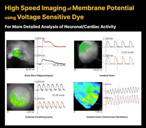
Voltage-sensitive dye changes its absorbance and fluorescence intensity when membrane potential changes in a stained brain or heart tissue.
By using voltage-sensitive dyes as chemical probes and capturing changes in light intensity with the use of a high-speed imaging device, it is possible to image in real time the activity of where, when, and how much excitation or inhibition occurred, in the brain and heart.
Product suitable for this kind of application
- High Speed Imaging System
- MiCAM03-N256
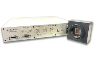
MiCAM03-N256 is a high-speed imaging system that captures and visualizes small changes in fluorescence intensity from biological samples stained with fluorescent probes, such as voltage sensitive dyes and calcium dyes.
- Spatial Resolution :
- 128x128 - 256x256 pixles
(32x32 pixels - 256x256 pixles with option) - Maximum Frame Rate :
- 1,000fps
(20,000 fps available with option) - Up to 2 camera heads can be used with completely synchronization.
- Ultra-High Sensitivity, Wide-Field Imaging System
- MiCAM03-C35IR
MiCAM03-C35IR is an imaging system equipped with a large 35mm full-size CMOS sensor, which has a spatial resolution of 2,160x1,280 pixels.
This model uses an ultra-high sensitive CMOS sensor which has large pixels, uses low noise technology, and has > 80% quantum efficiency. This makes it possible to image dim fluorescent samples at wide fields of view.
- Wide Field of View :
- 35mm full-size CMOS sensor
(47.7 mm diagonal) - Spatial Resolution :
- 2,160x1,280 pixels
- Maximum Frame Rate :
- 277.8 fps at 1,212x360 pixels